Augmented Reality and Virtual Reality market has a potential to reach 151 Billion USD with a paramount CAGR of 70.4% by 2022 (Source: Markets and Markets).
It is true that the AR and VR technologies have driven the gaming and entertainment industry, but it also has a very good potential to transform the healthcare industry since it actually can change a lot of traditional healthcare operations and branches in a number of ways, including radiology, oncology, training, and more.
Still being at a very early stage, digital reality (AR/VR) technologies are helping care delivery specialists to save life and take critical decisions. These technologies have a potential to bring overall healthcare cost down. Considering this potential of VR/AR, healthcare may revolutionize the way diagnostic practice is carried out to view MRI and CT images.
Let’s talk about diagnostic imaging in oncology. We know that medical imaging is crucial for all stages of oncology. Since FDA regulations focus on safety and effectiveness of devices, it has still not approved virtual reality or augmented reality viewing of medical imaging. However, there is an enormous potential for AR and VR applications in the medical imaging across its different stages namely detection, diagnosis and treatment.
Scope of Applying AR/VR in Medical Imaging
Considering the current advancement and maturity of VR/AR technologies, there are various ways in which these technologies can be applied to medical imaging operations:
- AR/VR can be used for tumor imaging during chemotherapy administration. Such AR/VR imaging applications can provide insights on changes in tumor size as well as other details such as end-to-end structure, shape, and margin. It can also help compare changes in the tumor over the period of time during chemotherapy.
- With current technologies in use, imaging provides two-dimensional information such as tumor location, laterality (in case of breast cancer), distance and size. With AR/VR applications, this information can be enhanced to deliver details with orientation, a 3D location of speculations and proximity to adjacent structures.
- With the use of AR/VR imaging, a detailed mapping can be done for pre-operative mark-up of tumor boundaries. This will help to understand the structure of a complex tumor. It will also help during lumpectomies for tumor resection and surgeons can make sure that no residual tumor is left.
Advantages of Utilizing AR/VR over Traditional Medical Imaging Techniques
There are some potential advantages with the use AR/VR compared most known volume rendering techniques in diagnostic imaging, which are listed below. These advantages are yet to be proven as no direct comparison has been performed for both these techniques.
- Volume rendering technique takes a lot more computer processing as it creates an artificial light source within the image to generate shadows for imaging. As it involves more computer processing, it has a higher possibility for interpretation errors related to processing. On the other hand, AR/VR imaging takes lesser computer processing to generate a three-dimensional imaging (as called the depth perception).
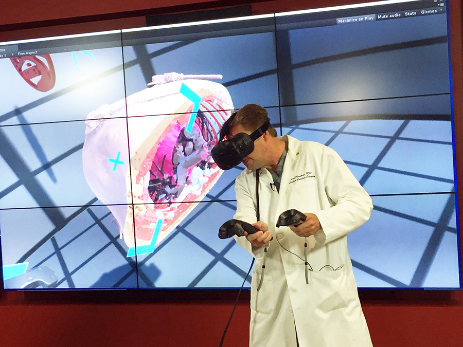
- The depth perception achieved from 3D imaging used with AR or VR may provide improved visualization and additional characteristic detail such as microcalcification (a tiny abnormal deposit of calcium salts) and its patterns such as compact clusters or a linear or branching pattern to determine the risk of cancer. Thus, AR/VR imaging may decrease the amount of manual thresholding by providing more details about complex structures.
-
- AR/VR environment may facilitate enhanced or additional digital content on radiology processing tools. An example could be, the use of different colors to represent different parts of a complex structure; use of different shapes like a cube or sphere (remember AR/VR provides 3D imaging) to represent similar anatomies.
- A radiologist, surgeon, and patient can improve their communication and collaboration with computer-generated graphics with this additional information. AR/VR enabled imaging may provide additional arrow or note(s) in imaging for communication at ease between aforesaid. It may also improve overall patient care procedures.
- Considering the advancements in Head Mounted Device (HMD), AR/VR devices can provide a better interface and control. Advanced head-mounted devices can track head motion and accordingly facilitate the virtual imagery. Such technology may allow a radiologist to deliver anatomical information quickly on virtual surface and help them to understand in details by providing the capability of zooming a particular part of the virtual image. Additional to zoom, they can change viewpoint positions, rotate the imagery, move the cursor, etc.
- As AR/VR provide 3D imaging and there is a possibility to deep dive with the capability of rotating, zooming and flying into a three-dimensional image with HMDs, there is a very high probability that we can detect tumor cells from the early stage which may remain unidentified with the current technologies and techniques.
- 3D augmented reality image may help a radiologist to deliver a clear picture of the structural anomalies and provides a more accurate diagnosis.
Augmented and virtual reality truly holds the future of medical imaging. This technology may bring the overall cost of healthcare operations down, reducing the possibilities of misdiagnosis and improve patient care activities.
eInfochips has hands-on engineering experience in engineering healthcare products for monitoring, diagnostics, analysis, and medical imaging. For more information, get in touch with us.


